Artificial muscles spring into action with mandrel-free fabrication technique
https://phys.org/news/2025-03-artificial-muscles-action-mandrel-free.html
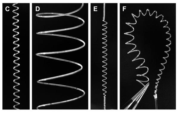
Phys.org · Artificial muscles spring into action with mandrel-free fabrication technique

Artificial muscles spring into action with mandrel-free fabrication technique
https://phys.org/news/2025-03-artificial-muscles-action-mandrel-free.html
 Biomimetic Origami: Planar Single-Vertex Multi-Crease Mechanism Design and Optimization
(Italian below)
We taking bets: children will quarrel over...
1. the truck/bus
2. the roof flat car
3. the roof fantasy car
4. the green car
---
Si accettano scommesse: i bambini litigheranno per...
1. il camion/autobus
2. l'auto con tetto piatto
3. l'auto con tetto a fantasia
4. l'auto verde
---
#STEM #STEMeducation #Mechanical @scuola @maupao
Great episode of #TechWontSaveUs with @timnitGebru
It's a real pleasure to listen to such a rich conversation on such diverse topics.
I especially liked how the topic of how the #AI industry labels people and methods was addressed.
It's the same for me, I've ended up assuming I'm a #DataScientist when I'm actually a #mechanical #engineer with a #PhD in #statistics. But the industry has decided that what I am is something I haven't studied about.
https://www.techwontsave.us/episode/267_ai_hype_enters_its_geopolitics_era_w_timnit_gebru
Is that a countdown timer, or just a clock?
Testing the lineart LoRa by @pixelworld_ai - sadly can't link it because it's no longer on CivitAI...
 "Electron spin dynamics guide cell motility"
https://arxiv.org/abs/2503.02923 #Physics.Bio-Ph #Mechanical #Dynamics #Quant-Ph #Q-Bio.Cb #Cell
"Investigation of Non-Radiative Relaxation Dynamics Under Pulsed Excitation Using Photon Absorption Remote Sensing: A Proof-of-Principle Study in Mechanical Sensing"
https://arxiv.org/abs/2502.19650 #Physics.App-Ph #Physics.Med-Ph #Physics.Optics #Mechanical #Dynamics #Matrix
The final #FreeCAD video is out! I cover one of the trickier subjects: finite element analysis. I use FreeCAD's FEM workbench to analyze deflection due to stress on a simple bracket.
https://youtu.be/FXG0xcjSV5Q
"Nonlinear contractile response of actomyosin active gels to control signals"
https://arxiv.org/abs/2502.18672 #Physics.Bio-Ph #Cond-Mat.Soft #Actomyosin #Mechanical
"ZnO@C/PVDF Electrospun Membrane as Piezoelectric Nanogenerator for Wearable Applications"
https://arxiv.org/abs/2502.16547 #Physics.App-Ph #Mechanical #Cell
"A highly sensitive, self-adhesive, biocompatible DLP 3D printed organohydrogel for flexible sensors and wearable devices"
https://arxiv.org/abs/2502.17208 #Cond-Mat.Mtrl-Sci #Physics.App-Ph #Mechanical #Adhesion
"Hierarchical poromechanical approach to investigate the impact of mechanical loading on human skin micro-circulation"
https://arxiv.org/abs/2502.17354 #Physics.App-Ph #Mechanical #Q-Bio.To #Cs.Ce #Cell
In case you missed it: my #FreeCAD assembly tutorial was released last week. Check it out if you want to learn about the new assembly workbench!
https://youtu.be/fMgSC9rcx_0
"Why epithelial cells collectively move against a traveling signal wave"
https://arxiv.org/abs/2008.12955 #Physics.Bio-Ph #Cond-Mat.Soft #Mechanical #Cell
Time for another #FreeCAD video! This one is probably the trickiest one yet: I cover fully organic shapes using the Curves workbench. It's definitely not intuitive, but it does work. Check it out here:
https://youtu.be/HDGdeLWGBns
"Nano-size fragmentation of Tantalum in Copper composite using additive manufacturing"
https://arxiv.org/abs/2502.04373 #Cond-Mat.Mtrl-Sci #Physics.App-Ph #Mechanical #Matrix